Newsroom
Science Update

Scientists develop a new gene therapy strategy for delaying aging
Recently, the researchers from the Institute of Stem Cell and Regeneration,CAS, Peking University, and Beijing Institute of Genomics, CAS have identified new human senescence-promoting genes by using a genome-wide CRISPR/Cas9 screening system and provide a new therapeutic approach for treating aging and aging-related pathologies. The study entitled “A genome-wide CRISPR-base screen identifies KAT7 as a driver of cellular senescence” was published online in Science Translational Medicine.
Jan 07, 2021
How many aging-promoting genes are there in the human genome? What are the molecular mechanisms by which these genes regulate aging? Can gene therapy alleviate individual aging? Recently, the researchers from the Chinese Academy of Sciences have shed new light on the regulation of aging.
Cellular senescence, a state of permanent growth arrest, has emerged as a hallmark and fundamental driver of organismal aging. Cellular senescence is regulated by both genetic and epigenetic factors. Despite of a few previously reported aging-associated genes, the identity and roles of additional genes involved in the regulation of human cellular aging remain to be elucidated. Yet, there is a lack of systematic investigation on the intervention of these genes to treat aging and aging-related diseases.
Recently, the researchers from the Institute of Stem Cell and Regeneration of the Chinese Academy of Sciences, Peking University, and Beijing Institute of Genomics of the Chinese Academy of Sciences have collaborated to identify new human senescence-promoting genes by using a genome-wide CRISPR/Cas9 screening system and provide a new therapeutic approach for treating aging and aging-related pathologies. The study entitled “A genome-wide CRISPR-base screen identifies KAT7 as a driver of cellular senescence” was published online in Science Translational Medicine on January 6th, 2021.
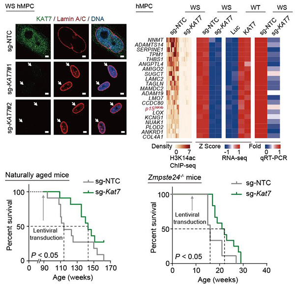
In this study, the researchers conducted genome-wide CRISPR/Cas9-based screens in human premature aging stem cells and identified more than 100 candidate senescence-promoting genes (Figure 1). The researchers further verified the effectiveness of inactivating each of the top 50 candidate genes in promoting cellular rejuvenation using targeted sgRNAs. Among them, KAT7 encoding a histone acetyltransferase, was identified as one of the top targets in alleviating cellular senescence. KAT7 increased in human mesenchymal precursor cells during physiological and pathological aging. KAT7 depletion attenuated cellular senescence whereas KAT7 overexpression accelerated cellular senescence. Mechanistically, inactivation of KAT7 decreased histone H3 lysine 14 acetylation, repressed p15INK4b transcription, and rejuvenated senescent human stem cells (Figure 2).
Figure 1. A genome-wide CRISPR/Cas9-based screen for identifying human senescence-promoting genes.
Cumulative studies have described that age-associated accumulation of senescent cells and proinflammatory cells in tissues and organs contribute to the development and progression of aging as well as aging-related disorders. Prophylactic ablation of senescent cells mitigates tissue degeneration and extends the healthspan in mice. In this study, the researchers found that intravenous injection of a lentiviral vector encoding Cas9/sg-KAT7 reduced the proportions of senescent cells and proinflammatory cells in the liver, diminished circulatory senescence-associated secretory phenotype (SASP) factors in the serum, and extended healthspan and lifespan of aged mice. These results suggest that gene therapy based on single-factor inactivation may be sufficient to extend mouse lifespan (Figure 2). The researchers also found that the treatment with the lentiviral vector encoding Cas9/sg-KAT7 or a KAT7 inhibitor WM-3835 alleviated human hepatocyte senescence and reduced the expression of SASP genes, suggesting the possibility of applying these interventions in clinical settings.
Figure 2. Gene therapy targeting Kat7 extends lifespan in naturally aged and progeria mice.
Altogether, this study has successfully expanded the list of human senescence-promoting genes using CRISPR/Cas9 genome-wide screen and conceptually demonstrated that gene therapy based on single-factor inactivation is able to delay individual aging. This study not only deepens our understanding of aging mechanism but also provides new potential targets for aging interventions. (Contact:Guanghui Liu, ghliu@ioz.ac.cn)

Researchers reveal a unique cellular strategy in placenta to compensate nutrient deprivation during pregnancy
The research group led by Prof. Yang-Ling Wang at the Institute of Stem Cell and Regeneration,CAS, in collaboration with Dr. Yoel Sadovsky from University of Pittsburgh and Dr. Bin Cao from Xiamen University, have now revealed a unique strategy of STB to adapt to nutrient deprivation and thus support fetal survival. Their study was published in PNAS on Jan. 6, 2021.
Jan 07, 2021
During pregnancy, the health of the mother and the fetus is dominated by the appropriate allocation of nutrients between the two individuals. Maternal-fetal material exchange predominantly depends on the placenta, which plays critical roles in sensing fetal nutritional demand, modulating maternal supply, and adapting its nutrient transport capacity. Failures in the regulatory network of placental functions lead to serious clinical complications, such as preeclampsia, recurrent miscarriage, and fetal growth restriction (FGR), etc. FGR is defined as the pregnancy bearing a fetus that does not grow to full potential, largely due to insufficient delivery of maternal nutrition by the placenta. Annually, ~30 million newborns worldwide suffer from FGR, which leads to increased perinatal morbidity and mortality and multiple lifelong health problems.
Lining at the outer surface of the placental villi, the syncytiotrophoblast (STB) is directly bathed in maternal blood, and thus positioned to take charge of maternal-fetal exchanges of gases, nutrients, and waste. STB has been identified as the largest multinucleated epithelial surface in the body, which is formed through cell fusion of the mononucleated cytotrophoblast (CTB). Yet the advantages of such an extensively multinucleated cellular structure in substance exchange remains poorly understood.
The research group led by Prof. Yang-Ling Wang at the Institute of Stem Cell and Regeneration,CAS, in collaboration with Dr. Yoel Sadovsky from University of Pittsburgh and Dr. Bin Cao from Xiamen University, have now revealed a unique strategy of STB to adapt to nutrient deprivation and thus support fetal survival. Their study was published in PNAS on Jan. 6, 2021.
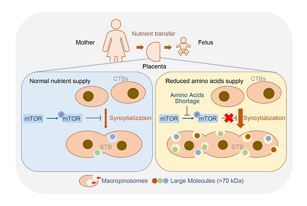
The researchers employed primary human trophoblasts and forskolin-exposed BeWo cells (a trophoblast cell line) as in vitro syncytialization models and found that macropinocytosis accompanied syncytialization in human trophoblast. Shortage of amino acids markedly promoted trophoblast syncytialization and macropinocytosis through repressing mammalian target of rapamycin (mTOR) signaling. As macropinocytosis is an efficient route to uptake fluid phase-derived large molecules by at least 10-fold, it empowers placental STB a developmental adaptation to ensure fetal demands when facing nutrient deficiency.
Consistently, the researchers treated pregnant mice with rapamycin, the inhibitor of the mTOR, to generate an animal model of FGR. The mouse placenta exhibited a greater extent of trophoblast syncytialization and enhanced macropinocytosis. Whereas pharmacologically blockage of macropinocytosis in these mice led to greatly worsened fetal growth due to the diminished macropinocytosis-dependent placental transport of large molecule.
The pathological relevance of macropinocytosis in pregnancy complication was revealed in FGR placentas which displayed notable repression in mTOR activity, augment in trophoblast syncytialization and macropinocytosis activity.
Taken together, this study discovered that differentiation of trophoblasts toward syncytium triggers an endocytosis strategy, macropinocytosis, to uptake large extracellular molecules. This unique machinery of nutrient uptake is strikingly boosted via inhibition of mTOR under amino acid shortage conditions, which is essential for fetal survival. The findings underscore a novel and physiologically important compensatory pathway for placental multinucleated trophoblasts to achieve a prime goal of nutrient delivery in the context of poor blood flow or other limitations in maternal nutrient supply. The study may facilitate the development of potential treatments to support fetus that suffers from maternal malnutrition or other conditions leading to insufficient nutritional transport.
The working model of macropinocytosis-mediated adaptation to nutritional stress in placental syncytiotrophoblasts.
Contact
Yan-Ling Wang
Institute of Zoology
E-mail: wangyl@ioz.ac.cn
http://english.rpb.ioz.cas.cn/groups/wangyanling/
Reference
Placental trophoblast syncytialization potentiates macropinocytosis via mTOR signaling to adapt to reduced amino acid supply
https://www.pnas.org/content/118/3/e2017092118

